Key takeaways
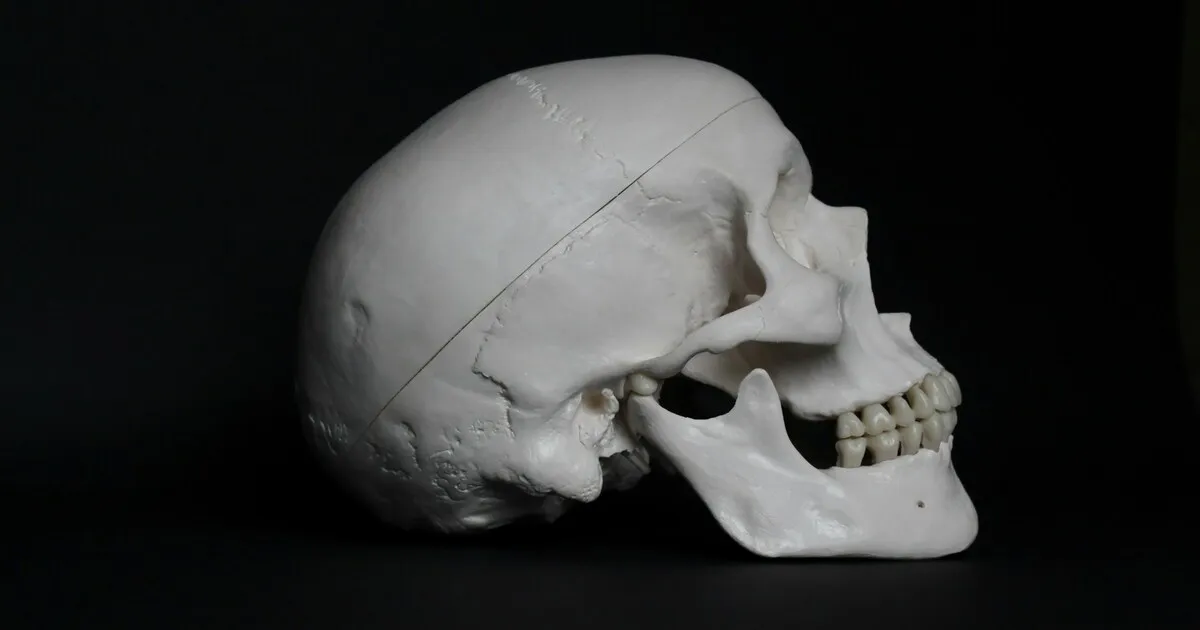
- Short face syndrome is a skeletal pattern: the lower third of the face is too short for the rest of the face. Typical signs are little or no upper tooth display at rest, a deep bite, a strong chin, a deep fold under the lower lip and a face that can look older or "overclosed".
- When the upper jaw is too short vertically, the condition is called vertical maxillary deficiency, and the operation that treats it is a maxillary downgraft: a Le Fort I osteotomy that moves the upper jaw down.
- Moving the upper jaw down is among the least stable movements in jaw surgery. Interpositional grafting, heavy rigid fixation, operating on the lower jaw at the same time and patient-specific plates designed on a 3D plan are used to limit relapse, but some vertical relapse can still occur.
- A downgraft often travels with lower jaw surgery. Because the lower jaw rotates down and back with it, and because moving both jaws makes the downgraft more stable, short face correction is usually planned as bimax (double jaw) surgery, often with a genioplasty.
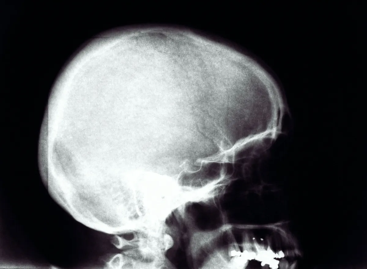
- The diagnosis is made on a full CT scan, not on photographs: worn teeth can mimic a short face and need a completely different treatment.
Most writing about jaw surgery is about faces that are too long, too convex or too prominent. Short face syndrome is the quieter opposite, and it is under-diagnosed for a simple reason: a short face rarely looks "wrong" to its owner. It looks strong, square and closed. What people notice instead is that they never show their upper teeth when they talk, that their lips look thin, that a deep crease sits under the lower lip, or that their face looks older than it is. This guide explains what short face syndrome is, how it is diagnosed, and how a maxillary downgraft, usually as part of bimaxillary surgery, gives the lower face back its height.
What is short face syndrome?
The term was defined in orthodontic literature in 1978, when Opdebeeck and Bell studied adults selected for a clinically reduced lower facial height and described the short face syndrome as a recognisable facial type with that reduced height as its common denominator. They also noticed that not all short faces are built the same way. In one group the lower jaw had a long ramus and a very flat mandibular plane. In the other, the posterior height of the upper jaw was sharply reduced: that second group is what surgeons call vertical maxillary deficiency.
The distinction matters because it decides the operation. If the upper jaw is short, lengthening the upper jaw is the logical treatment. If the upper jaw is normal and the problem sits in the lower jaw and chin, the plan looks different. Surgeons also describe the pattern as hypodivergent or brachyfacial, and a skeletal deep bite belongs to the same family; a deep bite on its own can also be purely dental.
How do you know if you have a short face?
There is no single measurement that defines short face syndrome, but the clinical picture is consistent. The table below compares the features surgeons look for.
| Feature | Balanced lower face | Short face pattern |
|---|---|---|
| Upper tooth display at rest, lips apart | A few millimetres of upper incisor visible | Little or none; the face can look toothless at rest |
| Bite | Normal overlap of upper and lower front teeth | Deep bite; lower front teeth hidden behind the upper ones |
| Lower lip and chin | Soft curve under the lower lip | Deep labiomental fold, everted lower lip, prominent chin |
| Jaw angles | Moderate | Often square, with strong chewing muscles |
| Lips | Visible red lip at rest | Lips look thin or rolled in, mouth looks "overclosed" |
| Overall impression | Proportionate | Strong but compressed; can look older than the age |
Many people with a short face come to a consultation asking for something else entirely: fuller lips, a lip lift, fillers in the lower face, or a "less aggressive" chin. Some of those treatments can help at the surface, but when the cause is skeletal, they are working against the bone underneath. That is why Dr. Cömert starts with proportions before any single feature:
"When the patient has a short face, I like to recommend bimax; but when they have a normal balance of the horizontal thirds of the face, usually I would go forward with facial implants or genioplasty."
Short face or worn teeth?
A lower face can also lose height because the teeth have worn down, been lost or been restored too short. That is a dental loss of vertical dimension, not a skeletal one, and it is treated by a dentist or prosthodontist rebuilding the teeth, not by moving the jaw. The two can look similar in a photograph. They look completely different on a CT scan, which is one reason the decision is never made from photos alone. In Dr. Cömert's words, "The final call is made on the CT scan, not on photos."
What is a maxillary downgraft?
The Le Fort I osteotomy is the standard way to move the upper jaw. Through an incision inside the mouth, above the teeth, the maxilla is separated from the rest of the skull along a horizontal line placed safely above the roots of the teeth. Once it is free, it can be moved forward, backward, up, down or rotated. Moving it down is what surgeons call a downgraft, an inferior repositioning or a maxillary lengthening.
Why the gap needs a graft
When the upper jaw moves up, bone sits on bone and heals quickly. When it moves down, a gap opens between the jaw and the midface above it. Chewing forces push upwards on the teeth every day, and without support the jaw tends to drift back towards where it came from. This is why, in the widely cited hierarchy of stability published by Proffit, Turvey and Phillips, downward movement of the maxilla sits in the "problematic" group: without special fixation, up to half of patients showed more than 2 mm of change in the first year.
Surgeons answer this in several ways. An interpositional graft, bone or a bone substitute, fills the gap so the jaw rests on something solid. Rigid fixation, heavier than for other movements, holds the position while it heals. Operating on the lower jaw at the same time helps too: Proffit and colleagues found the downgraft considerably more stable with a simultaneous ramus osteotomy, which reduces the biting force that pushes the healing upper jaw back up. And patient-specific plates designed on a virtual plan bring the jaw very close to the planned position: in one published case of total maxillary elongation, placement errors were under 1 mm at all seven landmarks measured.
Accuracy on the operating table is not the same as long-term stability. In a small 1989 series of 11 downgrafts of more than 5 mm, stabilised with plates and hydroxyapatite blocks, relapse was under 1 mm; larger datasets show more variable results, and even with rigid fixation some upward drift is common. Downward movement remains among the least forgiving movements in jaw surgery. The same applies to the counterclockwise rotation used for a recessed lower jaw, which also lowers the back of the upper jaw.
Why a downgraft is usually part of bimax surgery
The two jaws meet at the teeth, so they move as a pair. Lower the upper jaw and the lower jaw has to open slightly to keep the bite together, rotating clockwise around the jaw joints. The face gets longer, which is the goal, but the chin also moves back, which usually is not. In a short face that already has a strong chin this can be welcome to a degree; in a face with a normal or weak chin it needs to be compensated. There is also a stability reason: in the Proffit hierarchy, a downgraft is considerably more stable when the lower jaw is operated at the same time.
That is why short face correction is usually designed as bimaxillary surgery: a Le Fort I downgraft on the upper jaw, a bilateral sagittal split osteotomy (BSSO) on the lower jaw and, when the chin needs its own correction, a genioplasty. Surgeons call the three together a "trimax". Rotation of the whole maxillomandibular complex, clockwise or counterclockwise, is a well documented design, described by Wolford and colleagues for both function and appearance, that lets the surgeon change the angle of the bite as well as its height. In a 2025 retrospective series of 20 patients with short face syndrome and a square jaw, two-jaw surgery that lowered the upper jaw, with an inlay graft taken from the resected jaw angles, gave a mean gain of about 6 mm in total facial height at one year, with minimal relapse and no major complications.
The details of bimaxillary surgery itself, from the osteotomies to the orthodontic sequence, are covered in our complete guide to bimax surgery.
Do you need braces before a downgraft?
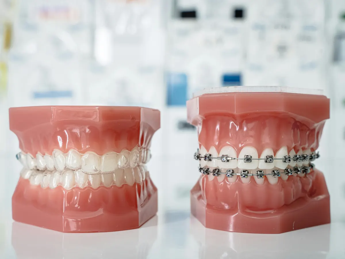
Traditional jaw surgery starts with a year or more of braces to prepare the bite. That is still the right path for some patients, but not for all. Dr. Cömert's position is clear: "If your teeth are aligned very well and the plan is a counterclockwise rotation, a downgraft or a slight advancement, orthognathic surgery can be done without any orthodontic treatment." In short face patients, though, a deep overbite and a deep curve of the lower teeth are common, so many need at least some orthodontic levelling; surgery-first is suitable only when the dental set-up allows it. When the teeth need work, a surgery-first approach is possible, with orthodontics continuing afterwards. Which pathway fits you is decided on your scans, not on a template.
How a downgraft is planned: custom jaw surgery
Because the downgraft is one of the movements with the least margin for error, it is where custom jaw surgery earns its value. In Dr. Cömert's practice every orthognathic case is planned digitally from CT data: the osteotomies are simulated, the movements are measured to the millimetre and patient-specific cutting guides and plates are prepared before the operation. The plates are not bent by hand during surgery; they are designed to hold the jaw in the planned position, gap included.
The amount is driven mainly by how much upper tooth shows at rest, a few millimetres being typical in young adults, and by the length of the upper lip; the front of the jaw is often lowered more than the back. Strong chewing muscles, common in short faces, are a known relapse risk and are part of the plan. In mild cases, a lengthening genioplasty, which adds height at the chin with an interpositional graft, can be an alternative to moving the upper jaw.
Two practical points patients often miss. First, the scan has to be the right one: Dr. Cömert asks for "the scan itself, not the radiology report", and a tooth-only CBCT is not enough to plan the whole face. Second, the plan is a conversation. How far the jaw moves is a balance between the face you want and the stability the bone can give, and as he puts it, "the movement wouldn't be too big. It has to stay within aesthetic limits."
What changes after a maxillary downgraft
- Upper tooth display. More of the upper teeth show at rest and in speech, which is often the change people notice first.
- Lower face height and lips. The lower third lengthens, the lips relax into a fuller, less compressed position and the deep crease under the lower lip softens.
- Bite. A deep bite can be corrected as part of the plan.
- Overall impression. The face can read as younger and softer, because a compressed lower face is one of the features we associate with age.
What a downgraft does not do on its own is change the soft tissue of the cheeks, the nose or the neck. Those are planned separately, and in some faces a patient-specific implant or a soft tissue procedure completes the result. Our guide to patient-specific facial implants explains when that is useful.
Recovery after downgraft and bimax surgery
The recovery follows the same pattern as any double jaw surgery in Istanbul:
- Days 1 to 2: hospital stay; swelling peaks around the third day.
- Weeks 1 to 2: rest at home; speaking and gentle activity return quickly.
- Weeks 1 to 6: a staged liquid to soft diet protects the healing bone. Our soft-food guide covers what to eat.
- Numbness of the upper lip, lower lip and chin is normal and fades over weeks to months.
- 6 to 12 months: the bone consolidates and the final facial balance settles.
International patients typically stay about 10 to 14 days in Istanbul for the first follow-ups and continue with remote check-ins using photos and video calls.
Risks and honest limits
A downgraft is a well established operation, but it carries the risks of any jaw surgery and one specific to its direction:
- Vertical relapse: the tendency of the jaw to move partly back up, reduced by grafting, rigid fixation and precise planning but not eliminated.
- Altered sensation in the lips, chin, gums or palate, usually temporary.
- Bite changes during healing that may need orthodontic fine tuning.
- Nose and lip changes: lowering the upper jaw can widen the base of the nose, change the position of the tip and alter the upper lip; a stitch to control nostril width is often used.
- Graft and sinus related problems, such as infection or congestion, and donor-site discomfort if your own bone is used.
- Bleeding, which is uncommon, and, rarely, a reduced blood supply to the upper jaw after large movements; jaw joint symptoms can also occur.
- Too much tooth or gum show if the movement is overdone, which is why the amount is planned around the upper lip, not around a target number.
Short face syndrome is uncommon compared with long face and retruded jaw patterns, and comparative studies are smaller. Choose a surgeon who will show you the plan in numbers and explain why the movement stops where it does.
Is a downgraft right for you?
If you recognise the pattern in this article, a short and strong lower face, little upper tooth display, a deep bite or a deep fold under the lip, the next step is not a decision but a diagnosis. Send front, profile and smile photographs, a photo of your bite and, if you have it, a full CT scan through the online assessment. Dr. Cömert reviews every jaw case personally and tells you whether the shortness is skeletal, dental or simply your normal anatomy, and what, if anything, he would change. If your concern is a recessed lower jaw instead, our article on mandibular retrognathia covers that pattern.
Prepared by the practice of Op. Dr. Mehmet Cömert, European Board-certified (FEBOPRAS) plastic, reconstructive and aesthetic surgeon, Istanbul. Quotations are from Dr. Cömert's own patient consultations. Last updated 24 October 2026. This article is general information and does not replace a personal medical assessment.